Earliest Parkinson’s Protein Clusters Seen in the Human Brain
October 8, 2025
For the first time, scientists have seen what they believe may be the earliest biological trigger of Parkinson’s disease inside the human brain. The discovery offers rare and direct evidence of how the condition might begin, long before symptoms appear, and could eventually help doctors detect and treat it much earlier.
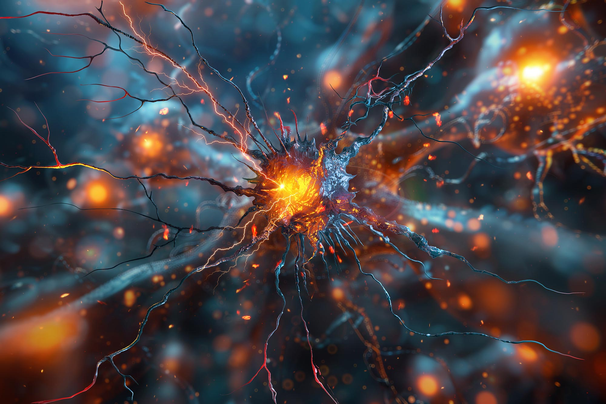
The research, led by teams from Cambridge, UCL, the Francis Crick Institute, and Montréal, used an advanced imaging method to examine donated human brain tissue. They focused on a protein called alpha-synuclein, which has long been linked to Parkinson’s. In healthy brains, alpha-synuclein helps nerve cells communicate. In Parkinson’s, however, the protein can misfold and clump together. These clumps, known as aggregates, are thought to disrupt brain function and kill nerve cells over time.
Until now, scientists could only see the large clumps known as Lewy bodies, which appear once the disease is well established. This new study, however, captured much smaller formations called oligomers. These are clusters of only a few misfolded proteins, and they are believed to form at the very start of the disease process. Detecting them directly in human tissue gives researchers a real glimpse of Parkinson’s in its earliest phase, something that had never been achieved before.
Using an ultra-sensitive version of fluorescence microscopy, the scientists were able to make these microscopic protein clusters visible. The technique enhanced the faint signals of the oligomers while suppressing background noise, creating clear, measurable images of where and how they appeared.
When comparing brain samples from people who had Parkinson’s with those from healthy people of similar ages, the difference was striking. Both groups had some alpha-synuclein oligomers, which means the presence of these clusters alone is not necessarily harmful. But the Parkinson’s brains contained far more of them, and they were larger and brighter, suggesting that the disease involves not just the formation of oligomers but also their abnormal growth and accumulation. The researchers also found a specific type of oligomer that existed only in Parkinson’s brains. This particular form may represent the spark that starts the disease long before symptoms emerge.
Understanding these early changes is crucial. Lewy bodies show where damage has already happened, but these smaller clusters might show where it begins. That difference matters because treatments developed to block or clear oligomers could, in theory, stop the disease from advancing before it causes serious damage. It could also make earlier diagnosis possible if scientists learn how to detect these protein changes in living people, perhaps through advanced scans or blood-based biomarkers.
Despite its significance, this discovery does not translate into a treatment yet. The research was conducted on post-mortem tissue, and the methods used to visualise these clusters cannot yet be applied in living patients. Parkinson’s is a complex condition with many possible contributing factors, and misfolded proteins are only one piece of the puzzle.
Still, this work marks an important turning point. For years, researchers have suspected that small, toxic protein clusters might be the true culprits behind Parkinson’s, while the larger Lewy bodies were more like the wreckage left behind. Now, for the first time, that suspicion has visual proof. Scientists can finally see the process they have theorised about for decades, and that opens a new path towards prevention and treatment.
For people living with Parkinson’s, it means progress is being made not only in managing symptoms but in uncovering what truly drives the disease. The hope is that one day, by targeting these early protein changes, Parkinson’s could be detected earlier, slowed down, or even stopped before it fully takes hold.
Comments (0)
Loading comments...