


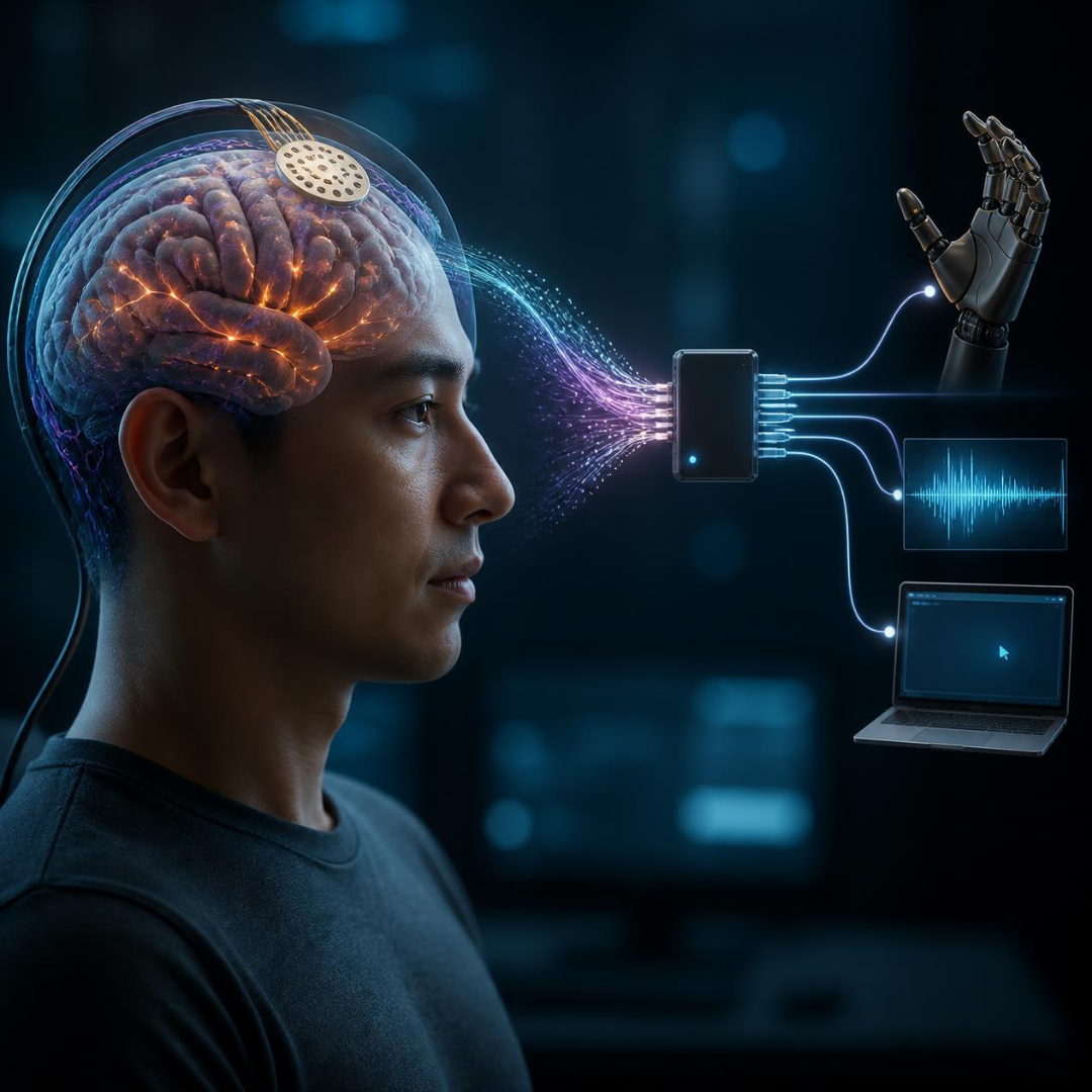
Surgical teams successfully place high tech brain sensors through micro slits to help restore movement and speech control
July 31, 2026
 Beatrice
Neuroscientists and engineers have built a tiny flexible sensor array that slides onto the surface of the brain through a paper-thin opening in the skull, opening up safer ways for people with Parkinson's to benefit from brain-computer devices without undergoing major open-skull surgery.
Brain-computer interfaces are microelectronic devices that listen to the electrical commands your brain sends out every second. When someone wants to move a hand or speak a word, millions of brain cells fire together, creating tiny electrical rhythms. Brain-computer devices pick up those faint electrical signals, clean them up using advanced computer software, and convert them into actions. For example, the software can turn a thought about moving an arm into actual movement by controlling a robotic limb, or it can translate thought patterns straight into words on a computer screen.
For years, getting a clear view of those brain signals required a compromise. If you put sensors outside on the scalp using a soft cap, the thick bone of the skull blocks most of the delicate electrical signals, making the readings fuzzy and limited. To get crisp signal clarity, surgeons traditionally had to perform an open-brain operation called a craniotomy. This meant removing a substantial section of the skull bone to lay down a flat sensor grid directly onto the brain, exposing a large area of delicate tissue to physical stress and longer recovery times.
This new technology changes that equation by using a tiny micro-slit in the skull instead of taking away a large piece of bone. Surgeons make a narrow cut, under a centimetre wide, and gently slide the device flat against the surface of the brain. The whole sensor structure sits on an ultra-thin, highly flexible strip of film that bends and flexes naturally with the surface folds of the brain. Because the film sits gently on top of the tissue without poking sharp pins into the brain itself, it causes far less irritation, reduces the chance of scarring, and remains safer over long periods.
Inside this thin film, engineers packed 1,024 individual sensor contacts into a tiny space. Each contact acts like a microscopic microphone, picking up local electrical chatter from nearby brain cells. Surgical teams tested the placement process using thin endoscopes, which are tiny cameras paired with precise guiding tools. In trials, putting the array safely in place took under twenty minutes, drastically reducing the time a person spends under anaesthesia. Multiple film strips can even slide through the same small opening to fan out and cover larger areas of the brain if needed.
When researchers analysed how well the system worked, the results proved exceptionally promising across several key areas:
The micro-slit approach removes the need for big surgical openings, which dramatically lowers the physical stress of surgery and speeds up recovery times.
The 1,024 sensor channels recorded exceptionally sharp electrical signals while subjects performed movement and language tasks, giving computers a clean feed of information to translate.
Tissue tests showed that the brain stayed healthy with no sign of severe inflammation or long-term damage, and the flexible arrays can be safely retrieved or replaced later without harming the underlying brain tissue.
Multiple sensor strips can connect through one small entrance point, allowing doctors to monitor broad networks across the brain without making larger cuts.
For people living with Parkinson's, these findings offer exciting long-term practical possibilities. The condition gradually disrupts the deep brain networks that control fluid movement, speech clarity, and timing, making everyday physical actions require intense effort. Having access to high-density sensor grids that slide into place with minimal surgical impact could streamline how future therapies are delivered.
Instead of relying solely on guesswork or external physical tests to monitor how well treatments work, a low-risk surface sensor could give doctors real-time data about electrical brain activity throughout the day. That exact information can help tailor medication timing or optimise deep brain stimulation settings with far greater precision. Additionally, as brain-computer interfaces continue to advance, these easy-to-place sensor arrays could help power assistive devices, helping restore smooth movement control and communication abilities without putting people through heavy open-brain surgery.
Comments (0)
Loading comments...