Brain Imaging is Mapping the Hidden Connections in Parkinson’s
April 7, 2026
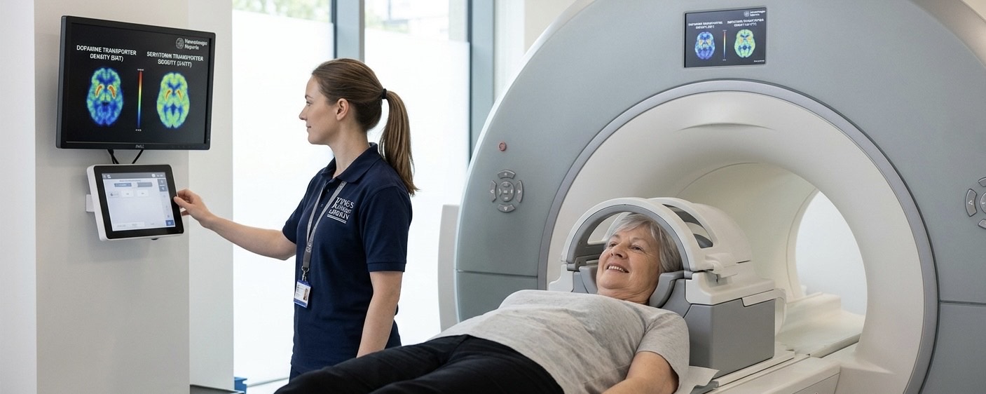
While Parkinson’s is traditionally identified by its motor symptoms, research has increasingly focused on the complex web of non-motor symptoms that often appear years before a diagnosis. A significant new study published in Neuroimage: Reports by researchers at King’s College London has used advanced molecular neuroimaging to reveal how the density of brain connections and different chemical systems—specifically dopamine and serotonin—relate to these various symptoms.
Beyond Dopamine: The Serotonin Connection
Dopamine has long been the primary focus in Parkinson’s research, particularly regarding motor control. However, this study underscores that the serotonergic system plays a vital role in many non-motor symptoms. By using Positron Emission Tomography (PET) scans, the researchers were able to measure synaptic density—essentially the number of connections between brain cells—and how these connections correlate with a person's experience of the condition.
The findings indicate that changes in the serotonergic system are deeply linked to mood-related symptoms such as anxiety and depression, as well as sleep disturbances. This suggests that the condition is truly a multisystem disorder, affecting more than just the brain's movement centres.
Mapping Motor and Non-Motor Symptoms:
The study looked at the association between brain markers and clinical symptoms in detail. Key takeaways include:
- Lower density of brain cell connections in specific regions was associated with both increased motor severity and a higher burden of non-motor symptoms. This measurement of synaptic density provides a global look at brain health rather than focusing on just one chemical.
- As expected, the loss of dopamine transporters was strongly tied to the "cardinal" motor signs—tremor, rigidity, and slowness of movement. This remains the hallmark of the physical challenges people face.
- Variations in serotonin transporters were more closely aligned with "hidden" symptoms. This includes the emotional well-being and sleep quality that significantly impact daily life but are often harder to quantify in a standard physical exam.
Why These Imaging Insights Matter:
Molecular imaging allows scientists to see objective changes in the brain's chemical machinery that clinical exams cannot detect. By understanding which specific systems are failing when certain symptoms appear, researchers can develop more targeted treatments. Instead of a one-size-fits-all approach focusing solely on dopamine, future therapies could be tailored to address the specific neurotransmitter imbalances unique to each person.
This research moves us closer to a future where we can predict the progression of the condition and intervene more effectively, improving the quality of life by addressing the full spectrum of Parkinson’s symptoms.
Comments (0)
Loading comments...