Parkinson’s Disease Associated With Neuroinflammation in the Brain
November 9, 2024
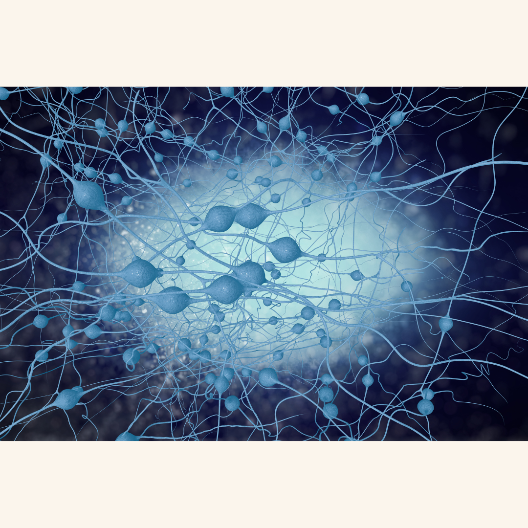
Yale scientists are using cutting-edge sequencing to study Parkinson’s disease in unprecedented detail. By analyzing cells in human brains affected by the disease, they’re learning more about how it develops. Their recent study, published on October 30 in Science Translational Medicine, mapped out different cell types in the brains of people with late-stage Parkinson’s. Comparing their findings with those from healthy brains and even brains affected by Alzheimer’s, they found distinct patterns in how Parkinson’s impacts brain cells, revealing an important role of inflammation in the disease.
This “cell atlas” study, led by Dr. Le Zhang, Dr. Sreeganga Chandra, and Dr. David Hafler, showed that Parkinson’s brains have higher levels of neuroinflammation, especially in two types of immune cells called T cells and microglia. This immune response may be a driving factor in the disease. The team also found that while Lewy bodies (clumps of protein found in Parkinson’s) accumulate in neurons, they actually correlate with lower levels of certain protective proteins. Boosting these proteins could be a future treatment approach to reduce harmful protein build-up.
When the team compared Parkinson’s to Alzheimer’s, they discovered that each disease affects neurons differently but shares inflammatory patterns in supporting brain cells. This finding shows how each disease has unique mechanisms but may share common pathways.
For future work, the researchers aim to study brain tissue from the early stages of Parkinson’s to better understand how it begins. They’re also investigating signs of inflammation in patients’ blood and spinal fluid, hoping to eventually design clinical trials to prevent Parkinson’s. This cell atlas is a crucial resource that researchers can use to continue exploring Parkinson’s and develop new therapies to manage it.
Comments (0)
Loading comments...