Retinal Imaging: A New Window Into Parkinson’s Progression
February 16, 2026
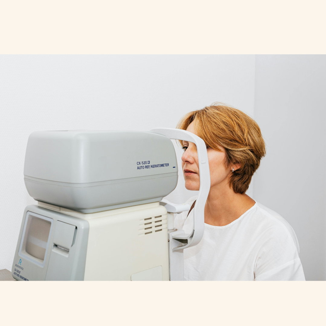
Research published in Scientific Reports has highlighted how simple, non-invasive eye scans could become a vital tool for monitoring the condition. By using Optical Coherence Tomography (OCT)—a common technology used by opticians—researchers found that changes in the thickness of the retina’s layers can accurately reflect the progression of the condition in the brain.
The Eye-Brain Connection
The retina is effectively an extension of the central nervous system. Because it contains the same types of neurons found in the brain, it acts as a "biological mirror." When neurons in the brain are affected by the condition, similar changes occur in the thin layers of tissue at the back of the eye.
Key Findings from the Research:
Specific Layer Thinning: The study identified that the inner plexiform layer (IPL) and the ganglion cell layer show significant thinning in people with the condition compared to those without it.
Correlation with Severity: The degree of thinning was found to correlate directly with the severity of motor symptoms. This means the thinner the retinal layers, the more advanced the condition typically is.
Early Detection Potential: Because these changes are detectable even in the early stages, retinal imaging could potentially identify the condition before significant physical symptoms appear.
Why This Matters for Management
The ability to use the eye as a proxy for the brain offers several advantages for both researchers and people living with the condition:
Non-Invasive Monitoring: Unlike lumbar punctures or expensive PET scans, an OCT scan takes only a few minutes, is completely painless, and is widely available at local opticians.
Tracking Treatment Success: In the future, these scans could be used in clinical trials to see if a new medication is successfully protecting neurons. If the retinal layers stop thinning, it’s a strong sign the drug is working.
Objective Data: For those managing the condition, this provides a clear, objective measurement of health that goes beyond subjective "how do you feel today" assessments.
Looking Ahead
This research moves us closer to a future where a routine eye appointment could provide critical data for a person's neurology team. By using the eye as a "window," clinicians can gain a clearer picture of what is happening in the brain, allowing for more personalised and proactive care.
Comments (0)
Loading comments...