


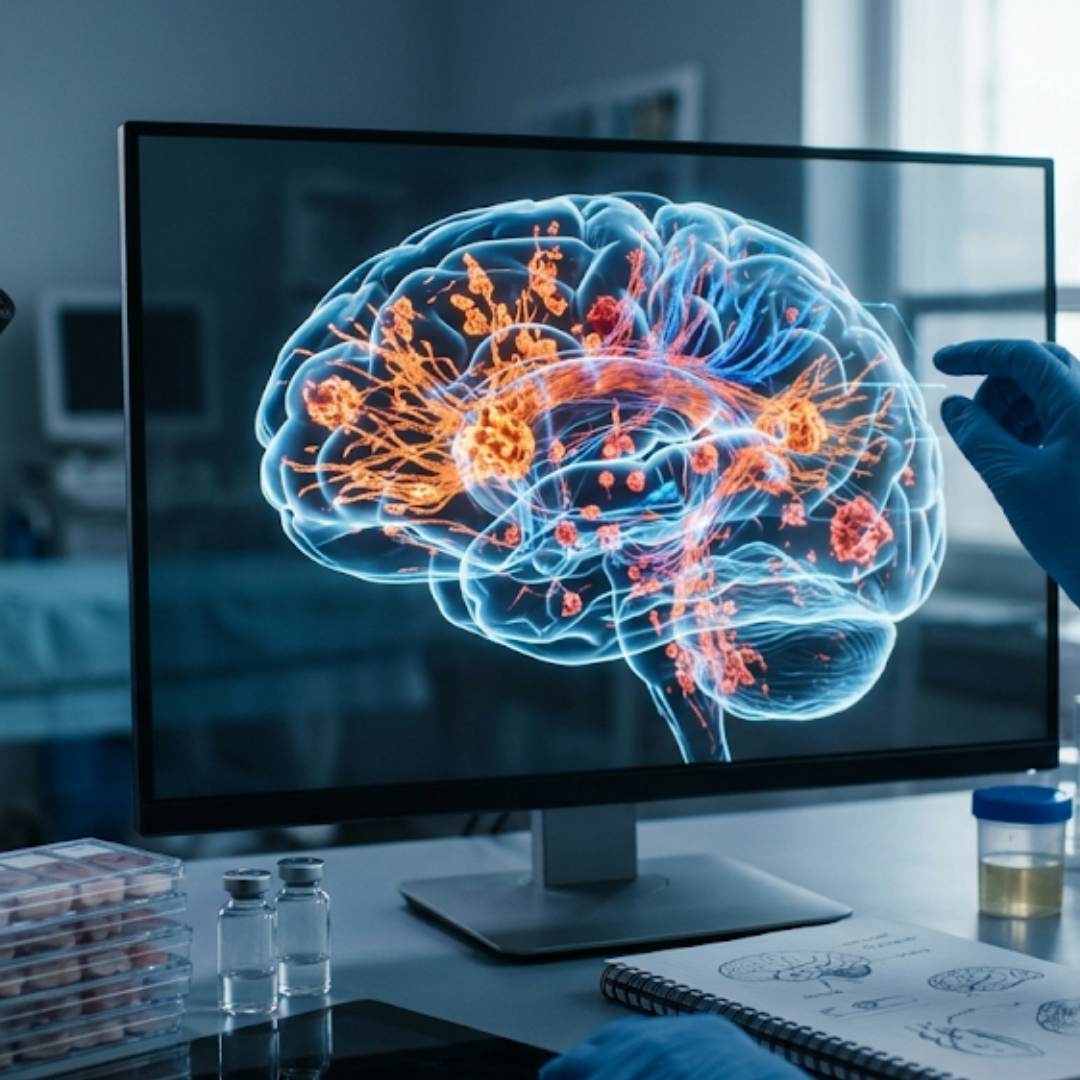
New brain bank study shows protein spread does not match Parkinson's progression
October 19, 2026
 Beatrice
For over twenty years, scientists have relied on a neat and tidy roadmap to explain how Parkinson's moves through the brain. Put forward by the German anatomist Heiko Braak in 2003, this famous theory suggests that clumped proteins called alpha synuclein start low down in the brainstem or the gut and slowly climb upwards into the higher regions of the brain like a rising tide. For a long time, researchers assumed that as these protein clumps spread from stage one to stage six under a microscope, a person's daily symptoms would worsen in direct, predictable lockstep.
A major October 2026 study published in the journal Neurology has put this long held assumption to the test and found that the microscope tells a very different story from real life. Led by Dr Lazzaro di Biase and Professor Alberto Espay alongside colleagues in Rome and Cincinnati, the research team examined records and donated brain tissue from sixty one people at the Parkinson's UK Brain Bank at Imperial College London.
The researchers wanted to see if a person's Braak stage at the end of their life actually matched how long they had lived with the condition, how old they were, or how much physical disability they experienced on the Hoehn and Yahr clinical scale. The results were striking in their complete lack of connection. Whether someone had protein clumps confined to the lower parts of the brain or spread widely across the outer cortex had no bearing on how severe their physical symptoms were. It also did not link to how many years they had lived with the condition or the overall weight of their brain. Someone could live with the condition for fifteen years and reach an advanced clinical stage while showing relatively limited protein spread, whereas another person might have widespread protein clumps with a very different clinical story.
What the researchers did find inside these donated brains was a noisy, crowded environment. Rather than seeing pure Parkinson's biology in isolation, the team discovered that overlapping age related changes were the norm rather than the exception. Over eighty per cent of the brains contained tau proteins, and nearly sixty per cent showed amyloid plaques, both of which are typically linked to Alzheimer's. Furthermore, sixty per cent showed signs of small blood vessel damage. Yet even the presence of these extra proteins and blood vessel changes did not vary across the different Braak stages.
Commenting on the paper, Professor Michael Okun noted that this work exposes the limitations of using pathology as a clock for clinical progression. He pointed out that Parkinson's is not one condition following one predictable path, and the frequent presence of other factors, including tau, amyloid, and vascular changes, further complicates the picture. For Professor Okun, the crucial takeaway is that scientists must be careful not to confuse what they see under the microscope with what people actually experience in their daily lives.
This disconnect matters enormously right now because the scientific world is racing to build new biological staging systems and biomarker tests designed to track the condition by measuring abnormal proteins in spinal fluid or blood. The authors of the UK Brain Bank study argue that simply looking at how much clumped protein has accumulated is not enough. Instead, researchers may also need to measure how much healthy, normal protein is left behind to do its job, alongside how many brain cells are actually surviving.
As Professor Okun summed up, as science develops new biological staging systems, biomarkers, and therapies aimed at slowing the condition down, researchers need to measure not only abnormal proteins but also nerve cell loss, daily physical impact, and the unique journeys of people living with Parkinson's. Ultimately, the person living with the condition, rather than the proteins under the microscope, must remain at the very centre of science.
Comments (0)
Loading comments...