Brain Maps Reveal How the Body Reorganises to Cope with Parkinson’s
February 12, 2026
While we often think of Parkinson’s as a condition that slows down the brain, new research suggests the brain is actually working overtime to compensate. A study published on the preprint server bioRxiv has revealed that the brains of people with the condition undergo a major "rewiring" process, strengthening connections in some areas to make up for weaknesses in others.
By comparing brain scans of people with the condition to neurotypical older adults, researchers at Rockefeller and Emory Universities have mapped out exactly how the motor system reorganises itself.
The Brain’s Backup System
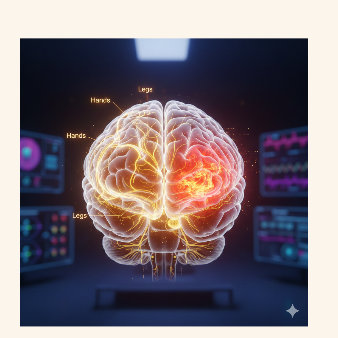
The study focused on "resting-state functional connectivity"—essentially, how different parts of the brain talk to each other while at rest. The researchers looked at the primary motor cortex, which controls different body parts like the hands, legs, and even the larynx (voice box).
They found that in people with Parkinson’s, the areas of the brain responsible for moving the legs and hands showed a significant increase in communication with the cerebellum. The cerebellum is the part of the brain that handles balance, coordination, and fine-tuning movement.
This suggests that when the primary pathways for movement start to struggle, the brain recruits the cerebellum as a "backup generator" to help maintain control.
Mapping Specific Struggles
The research didn't just look at movement as a whole; it broke it down by "effector" (specific body parts). This precision revealed some interesting patterns:
The Hand Connection: People in the study showed the greatest deficits in hand-related movements. Interestingly, the brain scans showed unique connectivity patterns for the hand area, suggesting the brain tries particularly hard to compensate for these complex tasks.
The Voice and Legs: Connectivity for the larynx and legs also showed significant changes, highlighting that the condition affects the entire motor map of the brain in distinct ways.
The Caudate Nucleus: This area, which is vital for starting movements, showed a mixed profile—it lost connection with some parts of the brain but gained it with others, showing the complexity of the internal struggle to keep the body moving.
Why This Matters for Therapy
Understanding these new "pathways" is about more than just curiosity; it provides a roadmap for better rehabilitation. If we know the brain is leaning on the cerebellum to keep the body moving, we can design therapies that specifically strengthen that relationship.
The researchers pointed out that this framework is particularly relevant for interventions like dance-based therapy. Because dance requires intense coordination, balance, and rhythm—all tasks managed by the cerebellum—it may be one of the best ways to train the brain’s compensatory circuits.
A New Framework
By moving away from looking at the brain as a single unit and instead focusing on specific motor subcircuits, this study offers a much more detailed picture of how Parkinson's impacts daily life. It confirms that the brain is remarkably resilient, constantly trying to find new ways to help people navigate the world despite the challenges of the condition.
Comments (0)
Loading comments...