Brain Stimulation Reveals Occipital Region Activity in Parkinson's Patients Diagnosed Within the Last 5 Years
October 1, 2024
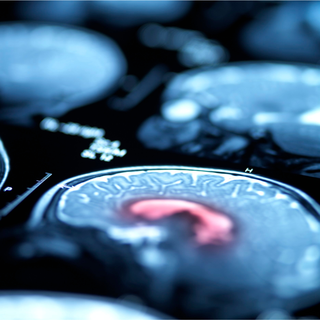
This study looked at how brain networks respond to external stimulation in people with Parkinson's disease (PD) compared to healthy individuals. Using a technique called TMS-EEG, researchers measured brain activity in different regions like the motor cortex, prefrontal cortex, and visual cortex in 62 Parkinson's patients and 76 healthy controls. The patients were divided into three subgroups: tremor dominant (TD), non-tremor dominant (NTD), and those with rapid disease progression (RDP).
The study found that Parkinson's patients showed weaker brain network connections, especially in the occipital (visual) region, compared to healthy people. This decrease in occipital network strength was more prominent in patients with faster disease progression. The results suggest that analyzing brain activity in this area could help identify different Parkinson's subtypes early on, offering insights for more personalized treatment. The study achieved 85% accuracy in identifying patients with rapid disease progression based on these brain activity patterns.
Comments (0)
Loading comments...